A groundbreaking preclinical study from Tufts University School of Dental Medicine and Tufts University School of Medicine introduces a novel concept that could transform the field of dental implantology: restoring proprioceptive feedback by avoiding traditional osseointegration.
Using a carefully designed rat model, the research team engineered titanium implants that interface directly with remaining periodontal nerve structures, challenging the conventional reliance on rigid bone-to-implant fusion.
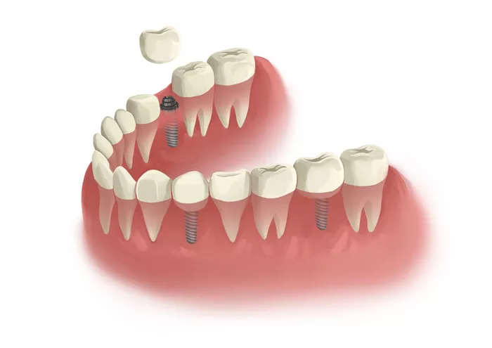
While dental implants have significantly advanced prosthodontics, they fall short of replicating the natural sensory function of teeth. Natural teeth are suspended by the periodontal ligament (PDL), which houses mechanoreceptors essential for proprioception. In contrast, conventional implants bond directly to the jawbone through osseointegration, eliminating this vital feedback system.
The absence of sensory input can impair fine motor control during chewing, swallowing, and other complex oral functions. To address this, the researchers developed a novel surgical method to extract mandibular incisors in brown Norway rats with minimal disruption. Custom-designed surgical blades allowed for precise dissection of the periodontal space, preserving residual neural elements within the socket.
Instead of relying on bone integration, the bioengineered titanium implants were press-fitted into the extraction sockets and sealed with a cyanoacrylate dressing. The implant surfaces were coated with biodegradable nanofibers infused with ultrastable fibroblast growth factor (FGF-β) and seeded with undifferentiated dental pulp stem cells. This coating was engineered to foster interaction with remaining nerve endings, particularly those associated with Ruffini corpuscles and the alveolar interface.
Weeks after implantation, clinical and radiographic evaluations showed that the implants remained stable, without signs of inflammation, infection, or displacement. Micro-CT imaging revealed a consistent radiolucent gap (0.7–0.9 mm) around the implants—evidence that osseointegration had not occurred. Importantly, the implants were not tender to pressure or percussion, suggesting a secure yet flexible attachment that may facilitate proprioceptive signaling.
The study’s authors propose that this nonrigid interface may enable neural integration at the implant-tissue boundary, potentially restoring aspects of tactile sensation and neuromuscular coordination absent in current implant designs.
Previous efforts to reintroduce sensory feedback in implants have largely failed due to the complete loss of periodontal nerves following extraction. This study takes a different approach by preserving terminal nerve endings and supporting their functional repair, rather than attempting full regeneration of the periodontal ligament.
By maintaining the biological integrity of the extraction site, the Tufts model supports partial preservation of the trigeminal nerve circuit, laying a foundation for future neural interface development in dental implants.
The selection of the rat model is also pivotal. Rodents such as brown Norway rats devote a substantial portion of their somatosensory cortex—up to 31%—to dental inputs, making them an ideal preclinical model for studying sensory restoration.
Despite the exciting potential of this approach, limitations remain. The small sample size and lack of direct neurodiagnostic evidence of sensory function restrict immediate clinical applicability. The researchers emphasize the need for advanced neuroimaging studies in future investigations to confirm proprioceptive restoration.
This innovative study opens new possibilities for developing bio-integrative implants that not only restore form and function but also reestablish the sensory experience of natural teeth.
Related topics:


